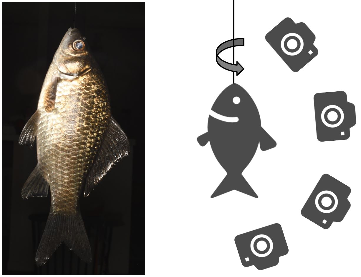

A simplified diagram of the photogrammetry method. The slowly rotating object (Carassius auratus langsdorfii), suspended by a nylon fishing line, is photographed from several angles. The resulting 3D model is displayed at https://skfb.ly/6ZRDz or https://ffish.asia/f/80064. — Kyushu University
Reporting in Research Ideas and Outcomes, a researcher from Kyushu University has developed a new technique to scan various plants and animals and reconstruct them into highly detailed 3D models. To date, over 1,400 templates have been made available online for public use.
Open any nature textbook or magazine and you’ll find stunning high-resolution images of the diversity of flora and fauna that encompass our world. From botanical illustrations in Dioscorides’ De materia medica (50-70 CE) to Robert Hooke’s sketches of the microscopic world in Micrographia (1665), scientists and artists have worked meticulously to depict the true majesty of nature.
The advent of photography has given us even more detailed images of animals and plants, large and small, in some cases providing new information about an organism’s morphology. As technology developed, digital libraries began to grow, giving us almost unlimited access to valuable data, with methods such as computed tomography or computed tomography and MRI becoming tools powerful for studying the internal structure of these creatures.
“Although powerful, MRI and CT scan methods are prohibitively expensive. You also cannot collect vital information such as the color of the organism,” says Yuichi Kano, associate professor of Kyushu University’s Graduate Education and Research Training Program in Decision Science for a sustainable society. “So we developed ‘bio-photogrammetry’ as a way to incorporate photogrammetry that could digitize and render a high-quality 3D image of an organism.”
Photogrammetry is a method by which you can obtain information and measurements about objects by analyzing photos or other images. Today, it’s commonly used to scan everything from landscapes to sculptures to create 3D digital models, similar to what you find on Google Earth.
Kano has used this same methodology to create thousands of models of various organisms.
“We hung the sample on a fishing line and took photos from multiple angles. We would end up taking hundreds of photos of the sample and feed up to 500 of the best ones into the photogrammetry program,” says Kano. “It’s similar to how the ‘bullet time’ sequences were filmed in the first Matrix movie, except instead of Keanu Reeves in a line surrounded by cameras, we use an octopus.”
While Kano has worked on various organisms including insects, plants, and even fungi, his current focus is on aquatic animals such as fish and amphibians. To date, over 1,400 specimens are available, all free, under the CC BY 4.0 license.
The current methodology has some limitations, such as the difficulty of capturing transparent creatures or creating extremely small models (<5 mm) or large (>1 m) organisms, but some improvements in software and protocols could help solve these problems.
“I hope to see this work continue to grow and be used in various fields like taxonomy, morphology and ecology. It’s free to the public, so you can use it for education or even plug it into a VR machine and explore these organisms up close. I’d love to see what some people can come up with,” Kano concludes.
For more information on this research, see “Bio-photogrammetry: digital archiving of colored 3D morphological data of creatures and associated challenges”, Yuichi Kano, Research Ideas and Outcomes (2022). https://doi.org/10.3897/rio.8.e86985
About Kyushu University
Kyushu University has been one of Japan’s leading research-oriented institutes of higher learning since its founding in 1911. Home to approximately 19,000 students and 8,000 faculty and staff, Kyushu’s world-class research centers Kyushu U cover a wide range of fields of study and research fields, from humanities and arts to engineering and medical sciences. Its multiple campuses, including the largest in Japan, are located around the city of Fukuoka, a coastal metropolis on the island of Kyushu in southwestern Japan, frequently ranked among the world’s most livable cities and historically known as a gateway to Asia.
Astrobiology
#Team #Tech #flora #fauna #fingertips #Astrobiology